


L. Bugeja1*, M. Vella2, K. Cortis3, T. Borg Barthet3, C. Cannataci3, S. Vella2, R. Sciberras2, C. Scicluna1, S. Chetcuti Zammit
1Department of Gastroenterology, Mater Dei Hospital, Malta
2Department of Medicine, Mater Dei Hospital, Malta
3Department of Radiology, Mater Dei Hospital, Malta
*Corresponding Author: L. Bugeja, Department of Gastroenterology, Mater Dei Hospital, Malta.
Abstract
Membranous obstruction of the inferior vena cava (MOIVC) is a rare disorder that had been classically thought to arise as a congenital malformation. However, it is now believed to be more commonly an acquired disorder. MOIVC may occur as a fibrous band or web formation secondary to thrombosis, whereby a fibrotic lamina replaces a regressed thrombus.
We report a 31-year-old gentleman presenting with hepatic failure and MOIVC. He was subsequently diagnosed with antiphospholipid syndrome, and the patient was managed with percutaneous balloon angioplasty, with average venous return via the IVC seen following the procedure.
Keywords: MOIVC, Anti-phospholipid syndrome, Liver failure, Interventional radiology
Introduction
Membranous obstruction of the inferior vena cava (MOIVC) is a rare disorder first described in 1909 in Japanese medical literature. [1] It was poorly defined until Okuda 1998 described MOIVC as separate from primary Budd-Chiari syndrome. [2] MOIVC is characterized by obstruction of the hepatic portion of the inferior vena cava (IVC) by either a membrane or a fibrous band and maybe a sequela to the occlusion of one or more of the hepatic veins.
MOIVC had been classically thought to arise as a congenital malformation. However, it is now believed to be more commonly an acquired disorder. MOIVC may occur as a fibrous band or web formation secondary to thrombosis, whereby a fibrotic lamina replaces a regressed thrombus.
MOIVC is described as having a chronic clinical course but can be complicated by an acute thrombosis and present as Budd Chiari Syndrome. We present a case of Budd Chiari syndrome secondary to MOIVC with associated non-occlusive thrombosis of the intrahepatic IVC in the context of a history of antiphospholipid syndrome and chronic alcoholism, treated with balloon angioplasty.
Case Presentation
A 31-year-old male with a medical history of antiphospholipid syndrome, chronic alcoholism, and substance misuse presented with a three-day history of fever, chills, rigors, and malaena. He exhibited signs of severe sepsis with systemic shock, acute renal failure, and liver failure, requiring intensive care unit admission for continuous hemodialysis and inotropic support.
Biochemical investigations demonstrated dual organ failure as evidenced by an acute kidney injury with severe metabolic acidosis, a creatinine of 609 umol/L and eGFR 10ml/min/1.73m2, along with liver synthetic failure, with a raised bilirubin level of 139.7 micromol/L and an International Normalised Ratio (INR) of 1.81. Inflammatory markers were increased by a white cell count of 15.66x10^9/L, CRP of 498.6mg/L, and pro-calcitonin level >100ng/ml.
An esophagogastroduodenoscopy showed grade 1 varices with no evidence of recent bleeding or red wale signs. A computerized tomography (CT) scan of the thorax, abdomen, and pelvis showed the portal vein reduced in caliber, with the hepatic artery and its branches leading to compensatory hyperdynamic enlargement, consistent with hepatomegaly flow.
There was also a significant narrowing of the supra-hepatic IVC, suggesting membranous obstruction of the inferior vena cava. An acute non-occlusive thrombosis was also observed in the infra-hepatic IVC. The azygos system was noted to be engorged with shunting of venous return below the diaphragm to the azygos arch.
Oesophageal varices, anterior abdominal wall collaterals, and subcutaneous collaterals were present. These findings were highly suggestive of membranous obstruction of the inferior vena cava, with the budd chiari syndrome and established cirrhosis with hepatomegaly flow.
An IVC angioplasty showed complete membranous obstruction of the portion of the IVC located between the liver and right atrium. This obstruction caused all the venous return of the abdomen and lower limbs to be channeled through the azygos and hemiazygos systems. After discussion at a local hepatobiliary multi-disciplinary team meeting, the patient underwent therapeutic venous balloon angioplasty of the IVC, and the occluded portion of the IVC was ballooned using two 12 mm diameter balloons, positioned side-by- side.
Post-procedure venography showed patency of the previously obliterated portion of the IVC with no flow of contrast via the azygos and hemiazygos systems - all venous return was noted to be transiting through the IVC into the right atrium.
A CT pulmonary angiogram (CTPA) was performed since the patient exhibited signs of respiratory distress despite optimal supportive treatment. The CTPA revealed a bilateral subsegmental pulmonary embolism with no evidence of right heart strain. After a repeat gastroscopy, he was anticoagulated with enoxaparin sodium to exclude actively bleeding varices or ulcers.
A raised anticardiolipin IgG >120U/ml, increased anti-b2 glycoprotein antibody >199 RU/mandlus positive lupus anticoagulant, confirmed antiphospholipid syndrome.
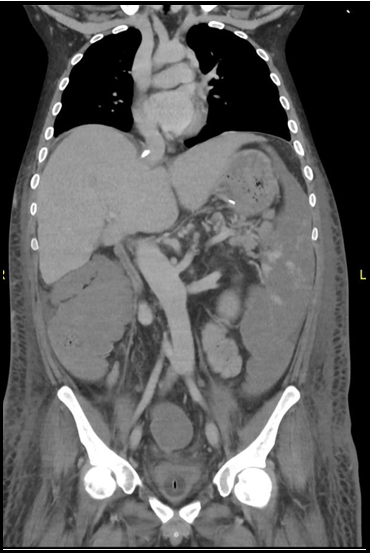
Figure 1: Coronal CT image in the portal venous phase showing a grossly enlarged spleen with areas of acute infarction. Note is also made of a calcification in the IVC at the level of the diaphragm with associated focal narrowing, compatible with chronic thrombosis. Signs of chronic liver disease are seen with an abnormal liver morphology. Subcutaneous oedema is alsonoted from the level of the diaphragm inferiorly.

Figure 2: Sagittal CT in the portal venous phase showing enlarged lumbar vein draining theinfraphrenic venous return into the azygos and hemiazygos system.
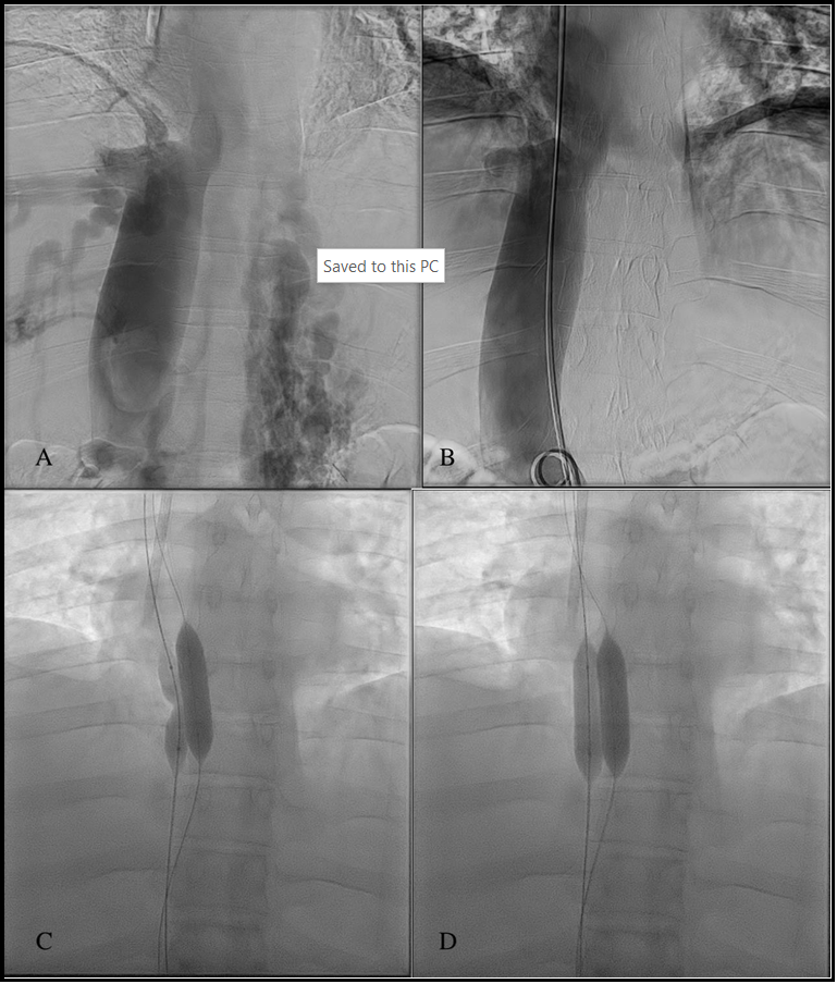
Figure 3: Venography pre and post venoplasty (a, b) using two balloons deployed side-by-side (c,d). No contrast is drain directly from the IVC to the right atrium prior to venoplasty. The venous return is seen to reach the right atrium via the azygos and hemiazygos systems. Normal venous return via the IVC is seen following venoplasty.
Discussion
MOIVC is a rare disorder first described in 1909 in Japanese medical literature. It was poorly defined until Okuda 1998 described MOIVC as separate from primary Budd-Chiari syndrome. MOIVC is characterized by obstruction of the hepatic portion of the IVC by either a membrane or a fibrous band and may occur in unison with the occlusion of one or more of the hepatic veins.
MOIVC may arise as a congenital malformation. However, it often appears as a fibrous band or web secondary to thrombosis, whereby a fibrotic lamina replaces a regressed thrombus. The thrombus may have formed secondary to an underlying hypercoagulable state, as in hematological disorders such as factor 5 Leiden mutation, an autoimmune disease such as antiphospholipid syndrome and systemic lupus erythematosus. MOIVC is generally described as having a chronic clinical course but can ultimately be complicated by acute thrombosis and presents as Budd Chiari Syndrome.
Such fibrous webs may lead to hepatic outflow obstruction, as in this patient, with venous return shunted to the azygos and hemiazygos venous systems. This patient also showed anterior abdominal wall collaterals draining pericardiophrenic veins on imaging that ultimately drained in mediastinal venous collaterals. Oesophageal varices were also evident, along with multiple subcutaneous collaterals throughout. This development of cavo-caval collateral anastomosis led to secondary liver cirrhosis in this patient, though his history of chronic alcoholism worsened his cirrhosis.
MOIVC may also be caused, apart from IVC webs, by thrombophlebitis of the IVC at an area close to hepatic vein outflow, which results in either stenosis of the IVC or complete obstruction; the literature available mentions Percival filariasis as a possible cause of thrombophlebitis, However, these rare cases are often seen in eastern countries such as Nepal and China.
Diagnosis of MOIVC is usually made through clinical examination and radiologically using ultrasound, computed tomography (CT), magnetic resonance imaging (MRI), and venography.
Management may include surgery or intervention, including membranotomies, shunts, radical resections, percutaneous transluminal angioplasty, and stenting. A liver transplant remains the optimal treatment choice for patients with liver failure.
Conclusion:
Membranous obstruction of the inferior vena cava with resultant hepatic outflow obstruction, hepatic venous congestion, and liver cirrhosis was treated with percutaneous balloon venoplasty. Average venous return via the IVC was seen following venoplasty.
Declarations:
Ethics approval and consent to participate: Not applicable.
Consent for publication: Written informed consent was gained from the patient’s next-of-kin as the patient passed away before writing up this report.
Availability of data and materials: Not applicable.
Competing interests: The authors declare that they have no conflict of interest.
Funding: This study was not supported by any funding.
Authors' contributions: The corresponding author drafted the manuscript. All authorsread and approved the final manuscript.
Acknowledgments: none.
References
-
Nagayo M (1909) Ueber die Obliteration der Hauptstaemme der Lebervenen und der unteren Hohlvenen in heparen Abschnitt derselben. Mittel Med. Fak. Keiser University Tokyo. 9: 1–13.