Qingping Joseph FENG1, George Shiyao HE2, Chun Peng GOH1, Ian James LONG1, Su Lone LIM1, Shiong Wen LOW1, Ira Siyang SUN1*
1Department of Neurosurgery, Ng Teng Fong General Hospital, National University Health System, Singapore
2Yong Loo Lin School of Medicine, National University of Singapore
*Corresponding Author: Ira Siyang SUN, Department of Neurosurgery, Ng Teng Fong General Hospital, National University Health System, Singapore.
Abstract
Background: Retropharyngeal kissing carotid arteries (KCA) is a rare anatomical variation of the common carotid arteries. Managing KCA in anterior spine surgery has rarely been reported in the literature. We detail the challenges of performing emergency anterior cervical discectomy and fusion (ACDF) in a patient with KCA and reviewed the current literature.
Case Description: A 67-year-old Female presented paraplegic after a fall. Pre-operative magnetic resonance imaging of the cervical spine demonstrated an acute prolapsed disc over the cervical three/ four (C3/C4) junction with cord signal change; KCA was also identified. She underwent ACDF surgery via the Smith-Robison technique. with the assistance of a vascular surgeon. The right carotid sheath was opened, its contents identified, isolated, and retracted medially. The remainder of the surgery was uneventful.
Conclusion: KCA presents as a technical challenge to anterior neck surgery. Early identification and collaboration with a vascular surgeon, is key to avoid serious complications.
Keywords: Kissing carotids, Anterior Cervical Discectomy and Fusion (ACDF), Anatomical variation, Multi-disciplinary tea
Introduction
Background
Retropharyngeal kissing carotid arteries (KCA) are a rare anatomical variation of the common carotid arteries (CCAs) [1]. It is characterized by the medial displacement of both vessels that appear to "kiss" each other either within the intrasphenoid or retropharyngeal space [2]. The development of KCA can be attributed to congenital anomalies involving the improper descent of the dorsal aortic root from the third branchial arch during the fifth to sixth embryonic weeks, leading to incomplete formation, accelerated linear growth and abnormalities [3]; or atherosclerotic changes or fibromuscular dysplasia which contribute to the medial displacement and tortuous caliber of the vessels [4]. Most patients with these types of variation are asymptomatic and findings are often incidental [4]. However, a small subset may still present with carotid bruit, dysphonia, dysphagia, pulsatile mass, hoarseness and symptoms of obstructive sleep apnea [4-6]. The presence of vessel tortuosity can lead to turbulent flow dynamics – this can accelerate the progression of atherosclerotic changes, and thereby increase the risk of cerebrovascular events [2,5,7,8].
KCA is also clinically significant due to the substantial risk they pose for catastrophic intraoperative complications, including severe and life-threatening bleeding as well as the potential for neurological deficits resulting from unintended vessel ligation and subsequent cerebral infarction [9]. While there are case reports documenting compilations related to otolaryngologic surgical procedures involving the upper pharynx, such as adenoidectomy, tonsillectomy, and uvulopharyngoplasty [2,3], there is a scarcity of reports in the English language literature addressing this rare phenomenon in cervical spine surgery [5,10-13]. This report aims to provide a review of the latest available literature on KCA, and to share our experience in effectively performing an anterior cervical spine surgery in a patient with KCA.
Case Description
We report a 67-year-old Female who presented with quadriplegia after tripping over a mattress, hitting her forehead and sustaining a hyperextension injury to her cervical spine. She reported spinal tenderness at the upper cervical region. Her sensory level was at cervical five, with reduced perianal sensation and anal tone.
Figure 1: T2-weighted MRI Cervical Spine. Sagittal (A) and axial (B) images. Thickened prevertebral soft tissue (A1) and signal change at the C3/C4 level seen in (A2). Retropharyngeal course of the CCAs (B1), and C3/C4 disc herniation (B2) demonstrated in (B).
An urgent Magnetic Resonance Imaging (MRI) of the cervical spine was obtained. This revealed a central disc herniation at cervical 3 /4 level (C3/C4), with T2-weighted/ Turbo Inversion Recovery Magnitude cord signal change. Other significant findings were of thickened prevertebral soft tissue and hematoma spanning from cervical 2 to cervical 6 levels, as well as anterior and posterior longitudinal ligament sprain injury. Of interest, a retropharyngeal course of the bilateral common carotid arteries was observed as highlighted in figure 1.
She underwent emergency Anterior Cervical Discectomy and Fusion (ACDF) surgery of the C3/C4 level which was performed with the assistance of a vascular surgeon. Awake nasal intubation was carried out with Aspen collar in-situ to minimize neck movement. She was placed in a supine position with head on donut pad, and neck in a neutral position. No neuromonitoring was used in view of her quadriplegic status.
After confirming the C3/4 level with intraoperative image intensifier, surgery was performed using Smith-Robinson approach. A right transverse skin-crease incision was made between the midline and the anterior border of the sternocleidomastoid (SCM). Platysma was cut followed by subplatysmal dissection. The medial border of SCM was identified laterally and strap muscles medially. This was followed by splitting the investing layer of deep cervical fascia.
The carotid pulse was palpable; however, the medial border of the carotid sheath could not be identified. The decision was made to open up the carotid sheath for direct visualization of its contents. With the common carotid artery identified, the medial border of the carotid sheath was easily recognized and subsequently retracted laterally along with its contents. Trachea, esophagus, and contralateral carotid sheath were also retracted medially. The vertebral body was palpated and the C3/4 disc space was confirmed again using image intensifier. The prevertebral fascia was then incised, followed by the undercutting of the bilateral longus colli muscles to accommodate placement of a self-retaining retractor. The anterior longitudinal ligament was found be disrupted with hematoma seen at C3/4 prevertebral space.
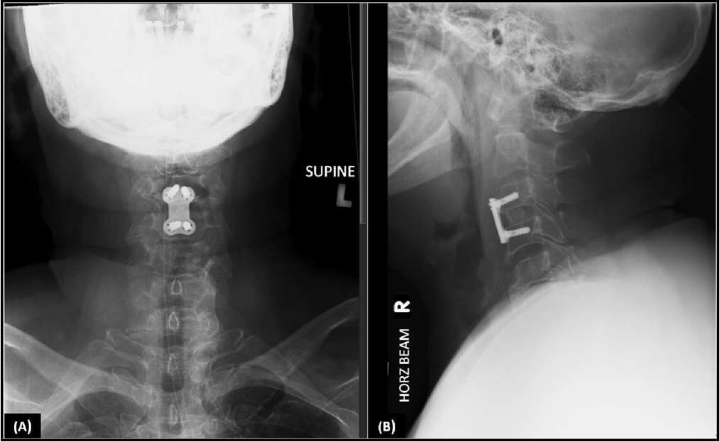
Under microscopic visualization, the ACDF proceeded in the usual manner. Autogenous iliac crest bone graft was harvested from the right iliac fossa, packed into the cage and secured with a Cervical Spine Locking Plate (Johnson and Johnson DePuy Synthes). Post- operatively, she was monitored in high dependency unit and was kept on Aspen collar for six weeks. X-rays of her cervical spine was obtained to look for satisfactory implant positioning, as shown in Figure 2.
Figure 2: X-ray Cervical Spine. Anterior-posterior (A) and lateral (B) views demonstrating the satisfactory positioning of the implants at the C3/C4 level.
Literature review
A comprehensive literature review was conducted on KCA. Titles and abstracts published up until 1st August 2024 were searched across the PubMed, Scopus, and Embase databases with the search terms "kissing carotid arteries" and "kissing carotids". Studies were included if they reported medially displaced retropharyngeal ICAs in human subjects. Articles were excluded if they: 1) were not available in a non-print English language format; 2) involved non-human subjects; 3) regarded non-surgical scenarios. This yielded 116 articles. An additional 7 studies were also included from reference lists of original articles set. After removing 10 duplicates, we carried out a title and abstract screening. 78 articles did not meet inclusion criteria, and an additional 7 articles were excluded due to irrelevance to surgical procedures. A total of 24 articles were identified for our literature review (see Appendix 1).
43 cases from 25 papers (including this paper) were reviewed. Patients ranged from pediatric to elderly and presented with heterogenous clinical and radiogram findings including prevertebral soft tissue swelling, progressive dysphagia, voice hoarseness and pharyngeal wall pulsations. A majority of cases were not related to operative management. Literature surrounding spinal surgery in the context of KCA is highlighted by 6 cases summarized in Table 2.
Table 1: Summary of patient characteristics, imaging and intervention
|
No. of studies |
Sex |
Average age |
Imaging |
Surgical Cases |
|||||
|
Male |
Female |
Unknown |
CT |
CT Angiogram |
MR Angiogram |
Spine |
Others |
||
|
25 (n=43) |
13 |
29 |
1 |
62.96 |
10 |
26 +2* |
5 +2* |
6 |
5 |
* 2 patients in 2 studies had both CT and MR angiogram done
Table 2: Summary of literature on KCA and spine surgery
|
No. |
Study |
Age |
Sex |
Presentation |
Imagi ng |
Type of Surgery |
|
1 |
Fix et al. 1996 |
64 |
F |
Severe cervical spondylosis, background previous laminectomy |
CT |
Modified ACDF to avoid carotid arteries |
|
2 |
Kumar et al. 2020 |
34 |
M |
Neck pain and progressive spastic quadriparesis with atlantoaxial dislocation |
CTA |
Posterior C1-C2 Decompression/ Fusion |
|
3 |
Mathkour et al. 2021 |
82 |
F |
Progressive cervical myelopathy |
CTA |
Combined Spine and ENT C3/C4 ACDF |
|
4 |
Inomata et al. 2023 |
69 |
F |
Progressive Cervical Myelopathy with irreducible atlantoaxial dislocation |
CTA |
Staged transoral Surgery |
|
5 |
Das et al. 2022 |
32 |
F |
Chronically progressive spastic quadriparesis with irreducible atlantoaxial dislocation |
CTA |
Posterior C1-C2 joint spacer placement and bilateral occipito- C3-C4 fixation |
|
6 |
This report |
67 |
F |
Paraplegia secondary to fall |
MRI |
Combined Spine and Vascular C3/C4 ACDF |
Discussion
We present a case report of a patient with aberrant anatomy of the common carotid arteries in the context of ACDF surgery. KCA is a rare congenital anomaly which significantly increase the complexity and risk of anterior cervical spine surgery. Review of the literature conclude that most cases are incidentally picked up on angiogram imaging. In our case, KCA was adequately identified on pre-operative MRI cervical spine.
An unanticipated encounter with such an anomaly during surgery can lead to catastrophic bleeding, stroke, or even death. The 6 spine cases were reviewed to look for operative considerations and eventual approach. 2 cases were modified from an anterior approach to a posterior-only approach to avoid the KCA entirely. Fix et al. reported the modification of transcervical approach for anterior cervical fusion to avoid vessel injury, but details of the surgical approach, management and outcomes were not discussed [12]. Inomata et al described a staged procedure, where they attempted to lengthen the carotid arteries through 1-week trial of halo traction [13]; this however resulted in no noticeable difference.
Our technique was closest to Mathkour et al, who described a combined operation with Otolaryngology [5]. A standard Smith- Robinson approach was utilized, with the additional steps of identification and displacement of the bilateral medial common carotid arteries. Their paper also advocated for pre-operative angiogram imaging, nerve monitoring, consideration of low-dose antiplatelet therapy and for electroencephalographic burst suppression to reduce cerebral metabolic demand.
The key learning point from our case is the importance of meticulous preoperative assessment of radiological images to avoid complications. Once the common carotid arteries (KCA) are identified, visualizing the contents of the carotid sheath is crucial for accurately recognizing its medial border. This technique is best performed by specialists with experience in vascular surgery.
Conclusion
Retropharyngeal kissing carotid arteries is a rare anatomical variation of common carotid arteries that present a unique challenge to anterior neck surgery. Knowledge of this entity, coupled with early and active identification of such an anomaly, even on routine or initial magnetic resonance imaging without accompanying angiographic images, is key in avoiding potentially devastating intra-operative and postoperative complications. This especially holds true in an emergent situation.
Authorship
QJF wrote the manuscript. GSH completed the literature review. CPG conceptualized the project and was part of the surgical team. IJL reviewed and edited the manuscript. SLL, SWL were part of the surgical team. ISS was the lead surgeon and oversaw the project.
Funding Sources: This paper was not supported by any sponsor.
References
- Windfuhr JP, Stow NW, Landis BN (2010) Beware of kissing carotids. ANZ journal of surgery. 80(9): 668-9.
- Paulsen F, Tillmann B, Christofides C, Richter W, Koebke J (2000) Curving and looping of the internal carotid artery in relation to the pharynx: frequency, embryology and clinical implications. Journal of anatomy. 197(Pt 3): 373-81.
- Ozgur Z, Celik S, Govsa F, Aktug H, Ozgur T (2007) A study of the course of the internal carotid artery in the parapharyngeal space and its clinical importance. Eur Arch Oto-Rhino- Laryngology. 264(12): 1483–9.
- Md Arepen SA, Abu Bakar AZ, Mohd Bakhit NHD, Muhammad Saifullah AAB, Salahuddin NA, et al. (2022) A Case Report of Kissing Carotid Arteries in the Retropharynx. Iranian journal of otorhinolaryngology. 34(122): 181–185.
- Mathkour M, Scullen T, Debakey M, Beighley A, Jawad B, et al. (2021) Anterior cervical discectomy and fusion in the setting of kissing carotids: A technical report and literature review. Clinical neurology and neurosurgery. 200: 106366.
- Van Abel KM, Carlson ML, Moore EJ (2013) Symptomatic internal carotid artery medialization: A rare anatomic variant resulting in cough, dysphonia, and dysphagia. Clinical Anatomy. 26(8): 966-970.
- Virvilis D, Koullias G, Labropoulos N (2013) Bilateral retroesophageal course of the carotid arteries. Journal of Vascular Surgery. 57(5): 1395-1397.
- Bissacco D, Domanin M, Schinco G, Gabrielli L (2016) Bovine Aortic Arch and Bilateral Retroesophageal Course of Common Carotid Arteries in a Symptomatic Patient. Vascular Specialist International. 32(3): 133.
- Garg A, Singh Y, Singh P, Goel G, Bhuyan S (2016) Carotid artery dissection following adenoidectomy. International Journal of Pediatric Otorhinolaryngology. 82: 98-101.
- Paulsen F, Tillmann B, Christofides C, Richter W, KOEBKE J (2000) Curving and looping of the internal carotid artery in relation to the pharynx: frequency, embryology and clinical implications. Journal of anatomy. 197(3): 373-381.
- Ozgur Z, Celik S, Govsa F, Aktug H, Ozgur T (2007) A study of the course of the internal carotid artery in the parapharyngeal space and its clinical importance. European archives of oto-rhino- laryngology. 264(12): 1483-1489.
- Fix TJ, Daffner RH, Deeb ZL, (1996) Carotid Transposition: Another Cause of Wide Retropharyngeal Soft Tissues. American Journal of Roentgenology. 167(5): 1035-1307.
- Inomata K, Takasawa E, Matsubayashi Y, Takayasu Y, Honda F, et al. (2023) Transoral Surgery for Irreducible Atlantoaxial Dislocation Complicated by Concomitant Aberrant Internal Carotid Arteries. Spine Surgery and Related Research. 7(2): 183- 187.


