


Joanna Wieckowska DO1*, Mark Wheeler DO1, Muhammad Aboudan MD1
1Ascension Genesys Hospital, Department of Pulmonary/Critical Care, 1 Genesys Parkway, Grand Blanc, MI 48439, USA.
*Corresponding Author: Joanna Wieckowska DO, 1Ascension Genesys Hospital, Department of Pulmonary/Critical Care, 1 Genesys Parkway, Grand Blanc, MI 48439, USA.
Introduction
Subcutaneous emphysema is the escape of air into the subcutaneous tissues, often secondary to injury to the parietal pleura from traumatic, surgical, infectious, or spontaneous etiologies [1]. Chest compressions during cardiopulmonary resuscitation (CPR) can be traumatic to patients and one of the most common thoracic complications sustained after CPR is rib fracture, which can lead to microscopic or macroscopic pleural injury [2,3]. Often when rib fractures are accompanied by subcutaneous emphysema, the clinician will suspect a more serious, life-threatening complication, such as pneumomediastinum, pneumothorax, or flail chest, that can lead to hemodynamic instability or collapse. Here, we present a case of CPR-related subcutaneous emphysema secondary to multiple rib fractures without other pulmonary complications.
Case Description
An 81-year-old male with a medical history significant for CML (in remission), prostate cancer s/p radiation, paroxysmal atrial fibrillation (on aspirin 81 mg daily), CAD s/p stent, CHF, ESRD on HD, presented to the hospital secondary to recurrent falls at home. The patient had tripped on his walker and fell to the ground, resulting in right 4th rib fracture, left 6th rib fracture, bruises, and abrasions. He was also found to have Gram-Negative bacteremia due to Pasteurella multocida and was put on Unasyn. He had concomitant weakness and dysphagia and ultimately required an NG tube to be placed for tube feeding. On day 10 of hospitalization, the patient’s NG tube required repositioning. At this time, his O2 demand began to increase, and he was placed on a 3L nasal cannula. Subsequent chest x-ray revealed appropriate NG tube placement but complete opacification of the left hemithorax. STAT CT Chest WO Contrast revealed complete atelectasis of the left lung and a soft tissue density in the left mainstem bronchus most compatible with mucous plugging. The patient was scheduled to undergo bronchoscopy the next morning, however, overnight he experienced PEA arrest. After 4 rounds of compressions, 3 doses of epinephrine, and 1 dose of sodium bicarbonate, the patient obtained a return of spontaneous circulation. Significant subcutaneous emphysema was clinically noted thereafter on the right lateral anterior chest wall, which was also seen on chest x-rays (Figure 1, 2).
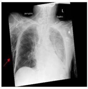
Figure 1: Extensive right lateral chest wall emphysema with no pneumothorax after ROSC.
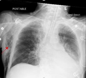
Figure 2: Moderate right lateral chest wall emphysema with no pneumothorax status-post one day after ROSC.
CT Chest WO Contrast the next day revealed moderate right chest wall emphysema with no pneumothorax, demonstration of fractures of right 4th and left 6th ribs with healing changes, new acute fractures of the right 1st, 2nd, 3rd, 5th, 6th, and 7th ribs, fractures with healing changes of the right 7th, 8th, 9th, and 10th ribs, minimally displaced fractures of the left 3rd, 4th, 5th, 6th, 7th, and 8th ribs, and fractures of the body of the sternum (Figure 3) along with continued obstruction of the left lower bronchus likely due to mucus secretion. Bronchoscopy was performed later that day and revealed a left lower lobe mucus plug, which was suctioned, and then left lung washing was sent for routine culture, AFB culture, fungal culture, cytology, and silver stain. Since being put on the ventilator and achieving ROSC four days ago, the subcutaneous emphysema gradually improved and ultimately resolved. Results of BAL revealed a positive bronchial culture with Enterobacter cloacae and Candida Albicans. The patient was started on Ciprofloxacin due to concern for CRE. After 9 days on the ventilator, the patient and the patient’s wife decided that the patient not be re-intubated after extubation. The patient was successfully weaned off the ventilator and extubated. Unfortunately, within 24 hours, the patient expired.

Figure 3: CT Chest Without Contrast after ROSC showing moderate right chest wall emphysema with no pneumothorax.
Discussion
In this specific case, the subcutaneous emphysema resulted from the multiple rib fractures status-post CPR. The rib fractures likely led to laceration of the visceral and parietal pleura and the adjacent lung parenchyma, causing the clinical and radiological findings. What makes this case remarkable is that despite the multiple rib fractures and subsequent subcutaneous emphysema there were no other overt pulmonary complications, such as pulmonary contusion, pneumomediastinum, pneumothorax, on either chest x-ray or non-contrast chest computed tomography, which is the more sensitive diagnostic modality for these diagnoses [4]. Subcutaneous emphysema usually heralds severe pleural or mediastinal injury [5].
The pathophysiology underlying its development entails alveolar overdistention which leads to rupture of peripheral pulmonary alveoli. Air then tracks up into the interstitial space to the mediastinum, where it may further extend to the pleural, peritoneal, or pericardial spaces, as well as the fascial planes of the neck and chest wall, resulting in subcutaneous emphysema [4,6]. In the setting of multiple, traumatic rib fractures, concern for more serious underlying pulmonary complications arise when subcutaneous emphysema appears. The number of rib fractures is also a strong predictor for more chest complications. Three or more rib fractures are found to be more sensitive to complications such as pulmonary contusion, pneumothorax, hemothorax, flail chest [7]. In one series, 81 % of patients with more than two rib fractures had either pneumothorax or hemothorax [8]. Despite a total of at least 12 rib fractures, a sternal fracture, and subcutaneous emphysema, our patient did not develop any other pulmonary complications during his hospital course with the subcutaneous emphysema ultimately resolving spontaneously.
References
- Kukuruza K, Aboeed A. Subcutaneous Emphysema. [Updated 2020 Aug 8]. In: StatPearls [Internet]. Treasure Island (FL): StatPearls Publishing; 2020 Jan-.
- Kralj E, Podbregar M, Kejžar, N, Balažic J (2015) Frequency and a number of resuscitation-related rib and sternum fractures are higher than generally considered. Resuscitation. 93: 136-141.
- Yusufoğlu K, Erdogan M, Tayfur I, Afacan AM, Çolak S (2018) CPR-related thoracic injuries: comparison of CPR guidelines between 2010 and 2015. Turk J Med Sci. 48(1): 24-27.
- Murayama S, Gibo S (2014) Spontaneous pneumomediastinum and Macklin affect: Overview and appearance on computed tomography. World journal of radiology. 6(11): 850–854.
- Liman ST, Kuzucu A, Tastepe AI, Ulasan GN, Topcu S (2003) Chest injury due to blunt trauma. European Journal of Cardio-Thoracic Surgery. 23(3): 374–378.
- Perraut M, Gilday D, Reed G (2008) Traumatic occurrence of chest wall tamponade secondary to subcutaneous emphysema. Cjem. 10(4): 387-391.
- Chien CY, Chen YH, Han ST, Blaney GN, Huang TS, et al. (2017) The number of displaced rib fractures is more predictive for complications in chest trauma patients. Scand J Trauma Resusc Emerg Med. 25, 19.
- Liman ST, Kuzucu A, Tastepe AI, Ulasan GN, Topcu S (2003) Chest injury due to blunt trauma. European Journal of Cardio- Thoracic Surgery. 23(3): 374-378.