


Ibrahim Touré1,3*, Youssouf Benmoh2,3*, Wissal Baghdad2,4, Sarah Slimani1,2,3, Houda Alloussi1,2,3, Amal Satté1,2,3, Ahmed Bourrazza1,2,3
1Department of Neurology, Mohammed V Military Teaching Hospital, Rabat, Morocco.
2Department of Neurophysiology, Mohammed V Military Teaching Hospital, Rabat, Morocco.
3Faculty of Medicine, Mohammed V University, Rabat, Morocco.
4Laboratory of Biology and Health, Neurosciences, Neuro-immunology and Behavior Unit, Faculty of Science, Ibn Tofail University, Kenitra, Morocco.
*Corresponding Author: Ibrahim Touré & Youssouf Benmoh, Department of Neurology, Mohammed V Military Teaching Hospital, Rabat, Morocco; Faculty of Medicine, Mohammed V University, Rabat, Morocco & Department of Neurophysiology, Mohammed V Military Teaching Hospital, Rabat, Morocco; Faculty of Medicine, Mohammed V University, Rabat, Morocco.
Abstract
Introduction
Eosinophilic granulomatosis with polyangiitis (GEPA), early known as Churg and Strauss syndrome, is a systemic necrotizing vasculitis of small- and medium-caliber arteries, associated with anti-neutrophil cytoplasm autoantibodies (ANCA). We report a case of GEPA revealed by acute multiple mononeuropathy.
Case Description
59 years old man, under medical therapies for arterial Hypertension, cortico-dependent late-onset asthma, admitted for asynchronous, asymmetric weakness with gait disorder, limb's burns evolving for five days in the context of altered general condition. Clinical examination revealed, areflexic distal tetra paresis. Electroneuromyography revealed axonal sensorimotor multiple mononeuropathy. Biological tests showed eosinophilic hyperleukocytosis above 10%, an inflammatory profile on plasma protein electrophoresis, and a positive myeloperoxidase (MPO) antibody. GEPA was diagnosed according to the clinical, biological, and radiological criteria defined by ACR/EULAR 2022 (American College of Rheumatology and European League Against Rheumatism). He was treated with methylprednisolone bolus combined to cyclophosphamide. The evolution was favorable with complete motor recovery and moderate paresthesia sequelae.
Conclusion
This is a rare condition that is difficult to diagnose in a neurology department due to the great heterogeneity of patients' clinical phenotypes. Extra-neurological manifestations essentially dominate initial symptoms. Neurological involvement may be the mode of revelation of the disease. Diagnosis is based on solid arguments defined by the ACR/EULAR 2022 criteria. Treatment with a combination of glucocorticoids and cyclophosphamide led to a marked improvement in peripheral neurological deficits and prolonged stabilization of the disease for ten months without recurrence.
Keywords: Eosinophilic Granulomatosis with Polyangiitis, Asthma, MPO-type Anti-ANCA antibodies, Hypereosinophilia, Acute Multiple Mononeuropathy.
Introduction
Eosinophilic granulomatosis with polyangiitis (EGPA) is a rare systemic vasculitis characterized by eosinophil-rich necrotizing inflammation of small- and medium-caliber vessels [1]. Clinical manifestations are diverse, with multi-organ involvement. The symptoms of GEPA are not specific, but their association leads to suspicion of the diagnosis [2]. A predominantly neurological clinical presentation highlights the diagnostic challenges of neurology training. We report a case of GEPA manifesting as acute, asynchronous painful weakness of the hands and feet, diagnosed and followed up in the course of our training.
Case Description
A 59 years old man, with pats medical history of arterial hypertension, with poor compliance, cortico-dependent late-onset asthma, on budesonide for 3 years. He was admitted seven days after the onset of symptoms marked by the sudden onset of weakness in the first three toes, followed by a deficit in wrist extension and grip in the right hand. Then, on the third day of the initial symptom, he reported the appearance of poorly systematized tingling-type paresthesia in the contralateral hand. On the fifth day of evolution, symptoms progressed to the lower limbs, with dorsiflexion weakness of the right foot, then of the contralateral side, without sphincter disturbance, or signs of respiratory distress or swallowing disorders. All these symptoms evolved in the context of altered general condition, with diffuse myalgias and a feverish sensation.
Clinical examination revealed a conscious, asthenic patient, hemodynamically and respiratory stable, supple neck, apyretic, well stained conjunctivae. Neurologic examination revealed bilateral steppage on unassisted walking, with right-hand drooping on Barre's maneuver (Figure 1), asymmetric distal flaccid tetraparesis, with abolished osteotendinous reflexes in the four limbs. The rest of the somatic examination (cardiac, pleuropulmonary, mucocutaneous, digestive, and osteoarticular) was normal.
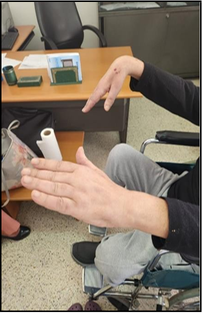
Figure 1: The patient's right hand droops.
Examination Of Segmental Muscle Strength
|
Upper Limb Muscular Strength. |
||
|
Muscle |
Left |
Right |
|
Finger Extensor |
2/5 |
4/5 |
|
Wrist Extensor |
2/5 |
4/5 |
|
Thumb Opponens |
4/5 |
4/5 |
|
Abductor Pollicis Brevis |
4/5 |
4/5 |
|
Finger Flexors (superficial/deep) |
4/5 |
4/5 |
|
Abductor Digiti Minimi |
4/5 |
5/5 |
|
Flexor Carpi Ulnaris |
2/5 |
4/5 |
|
Extensor Carpi Ulnaris |
2/5 |
4/5 |
|
Triceps |
5/5 |
5/5 |
|
Deltoid |
5/5 |
5/5 |
|
SCM / Trapezius |
5/5 |
5/5 |
|
Lower Limb Segmental Muscle Strength |
||
|
Muscle |
Right |
Left |
|
Flexor Hallucis Brevis |
4/5 |
3/5 |
|
Extensor Hallucis Brevis |
3/5 |
4/5 |
|
Tibialis Anterior |
3/5 |
3/5 |
|
Quadriceps |
5/5 |
5/5 |
|
Hamstrings |
5/5 |
5/5 |
|
Gluteal Muscles |
5/5 |
5/5 |
ENMG Findings (Table 1):
Table 1 : ENMG
|
Motor Conduction |
|||||
|
Nerves |
Distal latency |
Distal Amplitude (MV) |
Proximal Amplitude |
VCN (m/sec) |
F wave |
|
Median D |
NR |
NR |
NR |
NR |
NR |
|
Median G |
6.15 |
0.8 |
0.5 |
42 |
NR |
|
UInaire D |
NR |
NR |
NR |
NR |
NR |
|
UInaire G |
3.44 |
3.1 |
3 |
51 |
35 |
|
Peroneal D |
NR |
NR |
NR |
NR |
NR |
|
Peroneal G |
4.01 |
1.8 |
1.8 |
42 |
42 |
|
Tibial D |
NR |
NR |
NR |
NR |
NR |
|
Tibial G |
NR |
NR |
NR |
NR |
NR |
|
Sensitive Conduction |
|||
|
Nerfs |
Latence distale (msec) |
Amplitude distale (MV) |
VCS (m/sec) |
|
Median D III Palm |
NR |
NR |
NR |
|
Median G III Palm |
NR |
NR |
NR |
|
UInaire D |
NR |
NR |
NR |
|
UInaire G |
1.25 |
3.6 |
92 |
|
Sural D |
NR |
NR |
NR |
|
Sural G |
3.39 |
4.7 |
92 |
|
Peroneal superf D/G |
NR |
NR |
NR |
Axonal sensorimotor Multiple mononeuropathy, with denervation signs (fibrillation and positive slow wave on affected muscle needle detection).
Cerebrospinal fluid examination was normal.
CKemia and LDH were elevated (respectively 5104 IU/l and 494 U/L)
Blood cell count revealed hypereosinophelia (4600/ul). Biologic inflammatory profile was notified with high sedimentation rate (40 mm at first hour), elevated CRP and incipient beta-gamma block on serum protein electrophoresis. Infectious paraclinical tests were negative (Procalcitonin: 0.13 ng/ml; negative Serologies: HBV, HCV, TPHA, VDRL, PCR SARS COV2, urine examination).
Anti-ANCA MPO type was positive with elevated titer (99U/ml), anti-PR3 was negative. Proteinuria was negative.
Tumor markers: alpha-feto protein (AFP), carcinoembryonic antigen (CEA), and carbohydrate antigen (CA)-19-9; were normal.
Accessory salivary gland Histological examination was normal. Cerebral CT scan without contrast medium injection, troponin, electrocardiogram, and transthoracic cardiac echo-doppler were without abnormality.
Blondeau sinus radiograph (Figure 2) shows bilateral maxillary sinusitis.
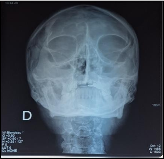
Figure 2: Sinus radiograph, Blondeau's incidence suggestive of bilateral maxillary sinusitis.
The diagnosis of eosinophilic granulomatosis with polyangiitis was made based on clinical, laboratory, and radiological findings according to the criteria defined by the ACR and EULAR 2022.
The patient underwent massive rehydration due to a hyperCKemia (26N), with quantification of diuresis and monitoring of weight, blood ionogram, and renal function. Combination treatment was instituted, comprising:
1. Methylprednisolone at a single dose of 1g/d for five days, then 1mg/kg/d for six weeks, gradually decreasing to the target dose of 20mg/d with adjuvant treatment for six months.
2. Cyclophosphamide IV at 600mg/m2 per infusion, the first three infusions at two-week intervals (D1, D15, D29) followed by three further infusions each 3 weeks. After six doses of cyclophosphamide, he was Started on oral azathioprine 150 mg/d with folic acid supplementation.
He underwent functional motor rehabilitation, with marked improvement in sensorimotor symptoms. Biological tests went normal after initial immunotherapy.
|
Muscle Strength Chart 45 Days Of Follow-Up |
||
|
Muscle |
Right |
Left |
|
Finger Extensor |
3/5 |
4/5 |
|
Wrist Extensor |
3/5 |
4/5 |
|
Thumb Opponens |
4/5 |
4/5 |
|
Abductor Pollicis Brevis |
4/5 |
4/5 |
|
Finger Flexors (superficial/deep) |
4/5 |
4/5 |
|
Abductor Digiti Minimi |
4/5 |
5/5 |
|
Flexor Carpi Ulnaris |
3/5 |
4/5 |
|
Extensor Carpi Ulnaris |
3/5 |
4/5 |
|
Triceps |
5/5 |
5/5 |
|
Deltoid |
5/5 |
5/5 |
|
SCM / Trapezius |
5/5 |
5/5 |
ENMG control realized after 6 months of follow-up showed Axonal sensorimotor multiple neuropathy with moderate electrical pattern amelioration.
After 10 months of follow-up, the neurological state was stationary, with no clinical relapse.
Discussion
GEPA belongs to a group of diseases characterized by necrotizing vasculitis affecting small and medium-sized systemic blood vessels associated with neutrophil anti-cytoplasmic cell antibodies ANCA [1, 2]. It is distinguished from other vasculitis by its association with late- onset asthma, peripheral eosinophilia, and upper respiratory involvement (nasal polyposis). Its annual incidence is estimated 1 to 3 per million adults. The average age of onset varies from 38 to 40, although cases have been diagnosed at ages ranging from 4 to 74, with no gender or race predominance [3].
We report a patient with GEPA, revealed by acute axonal sensorimotor multiple mononeuropathy according to ACR criteria.
The clinical manifestations of GEPA are protean, evolving in three phases. However, not all patients present all these phases, and they may intermingle during the disease.
The initial prodromal phase is dominated by general signs: malaise, fever, migratory polyarthralgia, myalgias and weight loss, respiratory discomfort. Myalgias and polyarthralgia have been reported in 37% to 57% of patients [3].
Upper respiratory symptoms such as chronic sinusitis (47%) and nasal polyposis (62%) are reported. Our patient presented malaise and neglected episodic polyarthralgia and bilateral maxillary sinusitis revealed by sinus radiography. The second phase is characterized by eosinophilic infiltration of target organs and peripheral eosinophilia. Cardiac involvement is observed in 62% of cases, but only 26% present clinical symptoms.
The clinical spectrum is diverse, including cardiomyopathy, arrhythmias, coronary artery disease, myocarditis, and pericardial effusion. Cardiac abnormalities are the main precipitating factors in intensive care units and are responsible for a third of EGPA-related deaths [1, 3]. Our patient had no cardiac involvement to the limit of troponin, transthoracic cardiac echo-doppler and ECG. Arterial hypertension is present in 10% of patients [3].
Respiratory and pulmonary disorders: late-onset asthma, which is virtually ubiquitous, is a key manifestation, occurring in 95-100% of patients, and may precede other systemic features for several years [2, 3]. In these patients, asthma is generally progressive and becomes corticosteroid-dependent in 75% of cases. Our patient had been treated for adult-onset asthma for three years. Diffuse alveolar hemorrhage is described in 3-4% of patients [3].
The third phase of the disease manifests itself 3 to 9 years after the onset of asthma and is characterized by neurological damage. Peripheral damage is the most common, accounting for 75-80% of GEPA-related cases [3, 4]. Multiple mononeuropathy is the classic presentation of peripheral disease in about 70-90% of patients [4]. The others showed distal symmetrical axonal polyneuropathy. Cases of acute polyradiculoneuropathy are increasingly described in the literature [1]. Cranial nerve damage is rare, but ischemic optic neuritis is the most frequently described cranial neuropathy [2, 3]. Damage to the central nervous system is rare but can lead to cerebral infarction or hemorrhage [5, 6]. Our patient presented with acute axonal sensorimotor multiple mononeuropathy with no cranial nerve involvement and no evidence of cardiac dysautonomia.
The third phase is associated with anti-neutrophil cytoplasmic antibodies (ANCA), with only 30-30% of GEPA patients being ANCA-positive. ANCA-positive patients directed against myeloperoxidase are the most common [6].
The diagnosis of GEPA is based on the clinical, biological, and histological features defined by the criteria adopted by the ACR and EULAR in 2022. These criteria have demonstrated a sensitivity of 85% and a specificity of 95% [3]. The diagnosis is accepted if the score is greater than or equal to 6 points. Our patient met the diagnostic criteria with a score of 9 points.
|
The ACR /EULAR Rating System In 2022 [7]. |
|
|
Criterion |
Score |
|
Maximum eosinophil count ≥ 10⁹ cells/L |
+5 |
|
Obstructive airway disease |
+3 |
|
Nasal polyposis |
+3 |
|
Positive ANCA or anti-proteinase antibodies |
-3 |
|
Extravascular inflammation with eosinophilic predominance |
+2 |
|
Mononeuritis multiplex or motor neuropathy not due to radiculopathy |
+1 |
|
Hematuria |
-1 |
The treatment protocol is diversified, mainly based on the severity and prognosis of GEPA, assessed by the five-factor score (FFS). The FFS score assesses the severity of GEPA and the impact of its factors on overall survival [8]. Each element counts for 1 point in the FFS score. The higher the score, the greater the impact on overall disease survival. Poor prognostic factors are as follows: Proteinuria > 1 gram/24h, creatinine > 440 µmol/l, specific cardiomyopathy, severe digestive involvement, and central nervous system involvement.
|
FFS (Five-Factor Score) |
Interpretation |
|
FFS = 0 |
None present |
|
FFS = 1 |
One of the five factors present |
|
FFS ≥ 2 |
At least two of the five factors present |
In 2009, the FFS score was revised to include two additional poor prognostic factors: age over 65 and absence of ENT manifestations [3, 8].
Glucocorticoid monotherapy was proposed for patients with an FFS=0 prognostic score. The combination of glucocorticoids and immunosuppressants is used for patients with a poor prognosis score ≥ 1. This combination therapy has also been proposed as a first-line treatment for patients with life-threatening manifestations, including eosinophilic alveolitis or severe peripheral neuropathy [6, 9].
Our patient had an initial FFS score = 0 with severe multiple mononeuropathy.
He received a combination therapy of methylprednisone and cyclophosphamide.
Our patient's clinical course was satisfactory, with significant recovery of the motor deficit and partial regression of the neuropathic pain. The evolution of GEPA under treatment could be impacted by the delay to initiate immunotherapy and the cardiac involvement. Overall survival at 10 years is of the order of 85%, subject to the absence of poor prognostic factors defined by the FFS score. [8]
Conclusion
This case illustrates that GEPA could be revealed by an essentially neurological clinical picture. GEPA remains an insidious disease with a protean phenotype involving neurological and extra-neurological manifestations. Diagnosis is based on the ACR /EULAR criteria.
The general management approach remains immunotherapy stratified by the FFS score. The combination of glucocorticoids and cyclophosphamide is indicated for GEPA with severe peripheral neuropathy.
Conflict of Interest: None.
Acknowledgement: None
References
- White J, Dubey S (2023) Eosinophilic granulomatosis with polyangiitis: A review. Autoimmun Rev. 22(1): 103219.
- Yoo IH, Choi ST, Choi SH, Kim JM, Ahn SW (2017) Eosinophilic Granulomatosis with Polyangiitis Presented as Acute Polyneuropathy and Cerebral Vasculitis. Exp Neurobiol. 26(3): 168-171.
- Chakraborty RK, Rout P (2025) Eosinophilic Granulomatosis With Polyangiitis (Churg-Strauss Syndrome). StatPearls Publishing.
- Khandelwal D, Singh M, Jagota R, Mathur V (2021) Eosinophilic granulomatosis with polyangiitis (Churg–Strauss syndrome) imitating Guillain–Barre syndrome (GBS): a case report. Egypt J Neurol Psychiatry Neurosurg. 57(1) : 173.
- Arsenijević M, Ivančević N, Jovanović D, Radović M, Berisavac I (2021) Rare case of recurrent stroke in a patient with eosinophilic granulomatosis with polyangiitis: a case report. Egypt J Neurol Psychiatry Neurosurg. 57(1): 76.
- Koike H, Nishi R, Ohyama K, Morozumi S, Kawagashira Y, et al. (2022) ANCA-Associated Vasculitic Neuropathies: A Review. Neurol Ther. 11(1): 21-38.
- Ponte C, Grayson PC, Robson JC, Suppiah R, Gribbons KB, et al. (2022) 2022 American College of Rheumatology/EULAR classification criteria for giant cell arteritis. Ann Rheum Dis. 81(12): 1647-1653.
- Beachy N, Satkowiak K, Gwathmey KG (2019) Vasculitic Neuropathies. Semin Neurol. 39(5): 608-619.
- Howarth N, Miller MA (2024) Sleep, Sleep Disorders, and Mental Health: A Narrative Review. Heart and Mind. 8(3): 146-158.